Life is a miracle in itself and the development of human life inside a womb is a magical process.
At the miraculous moment of fertilization – when the egg of a woman and the sperm of a man unite, a new human life begins. It will take roughly nine months for the baby to develop and be ready to be born from this point forward.
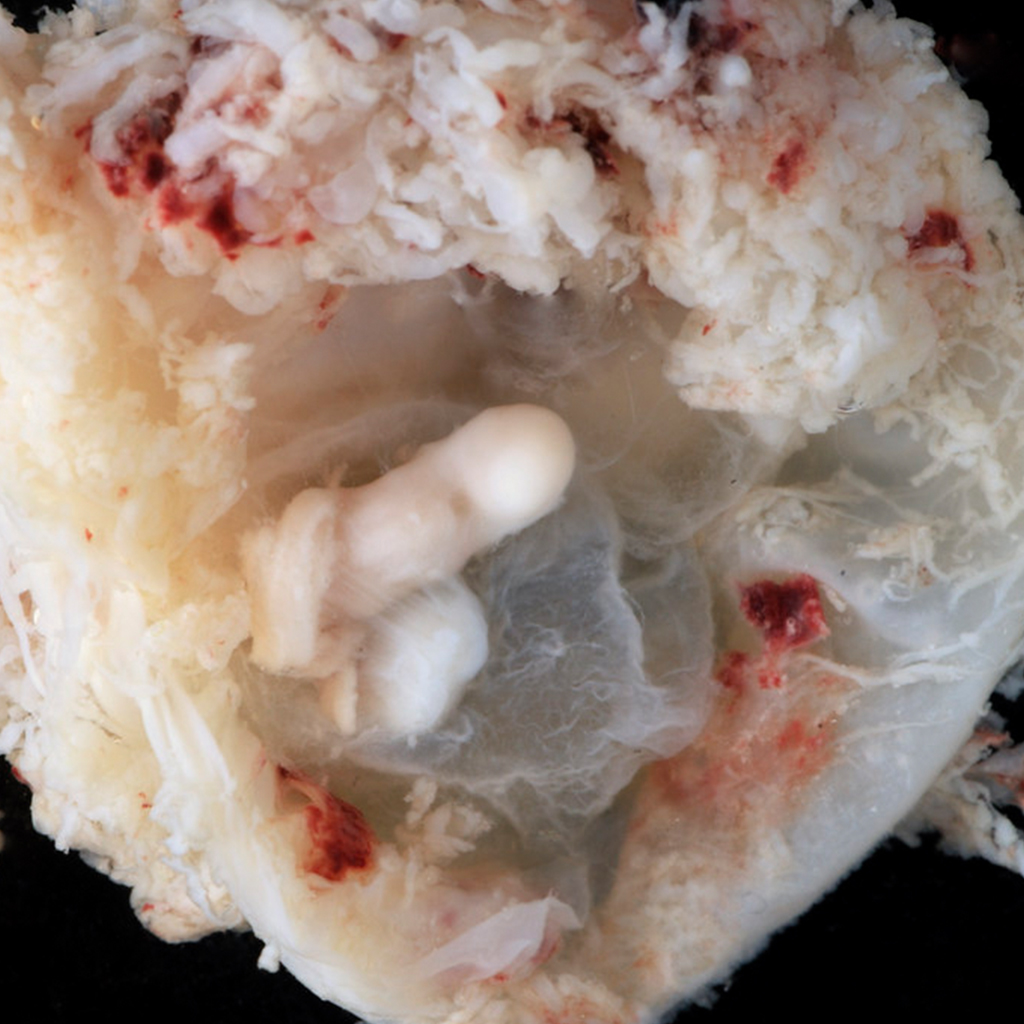
Embryo, 3 to 4 weeks. A 6mm embryo is identified which shows a possible heart bulge on the anterior surface. No limb bud is seen.
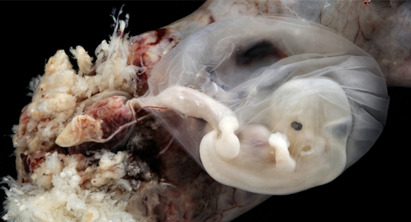
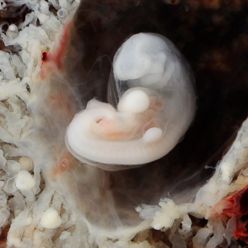
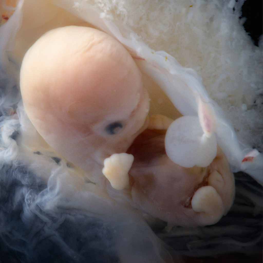
Embryo at 4 to 5 weeks. This is an embryo at approximately 4 to 5 weeks from conception. This was an ectopic pregnancy in a fallopian tube.
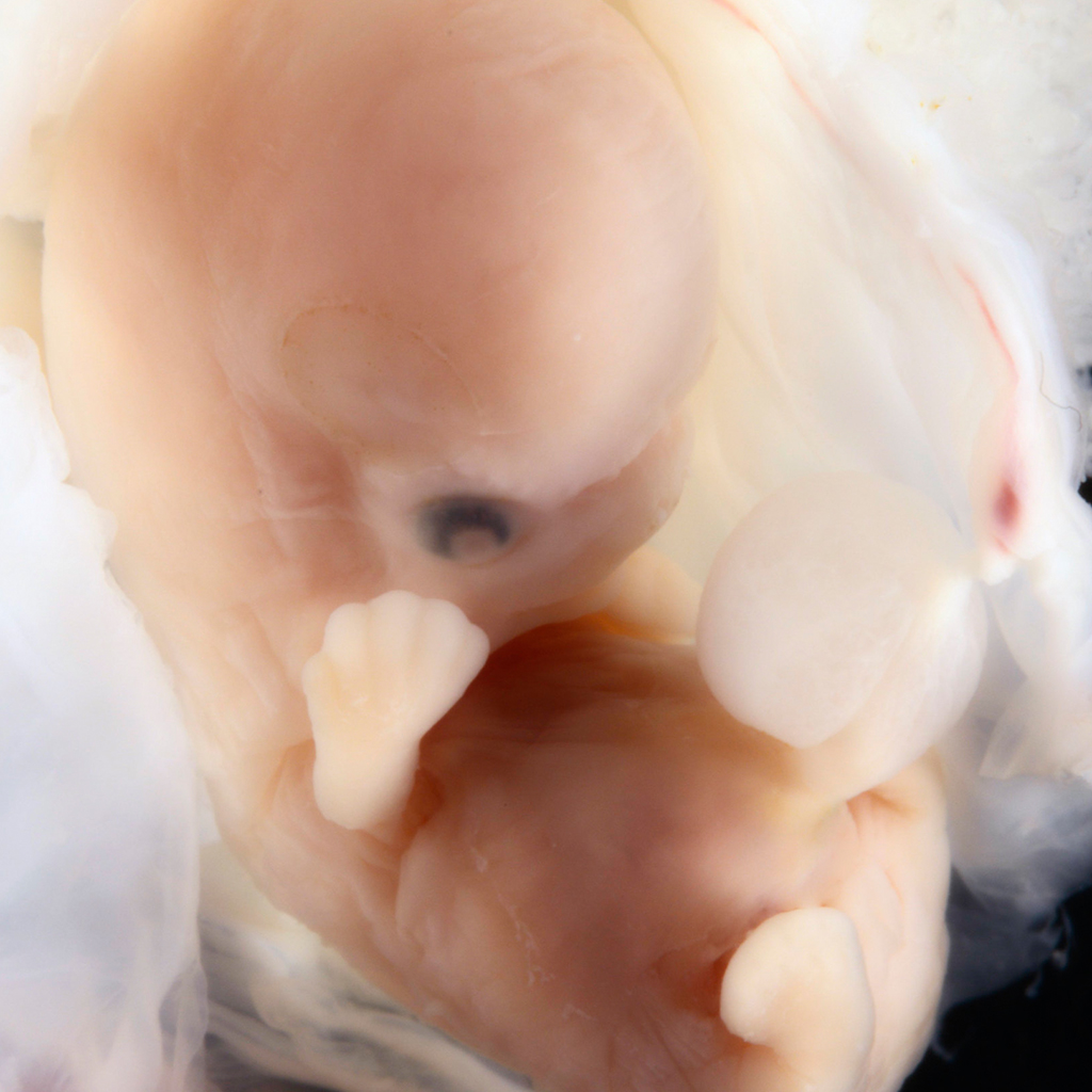
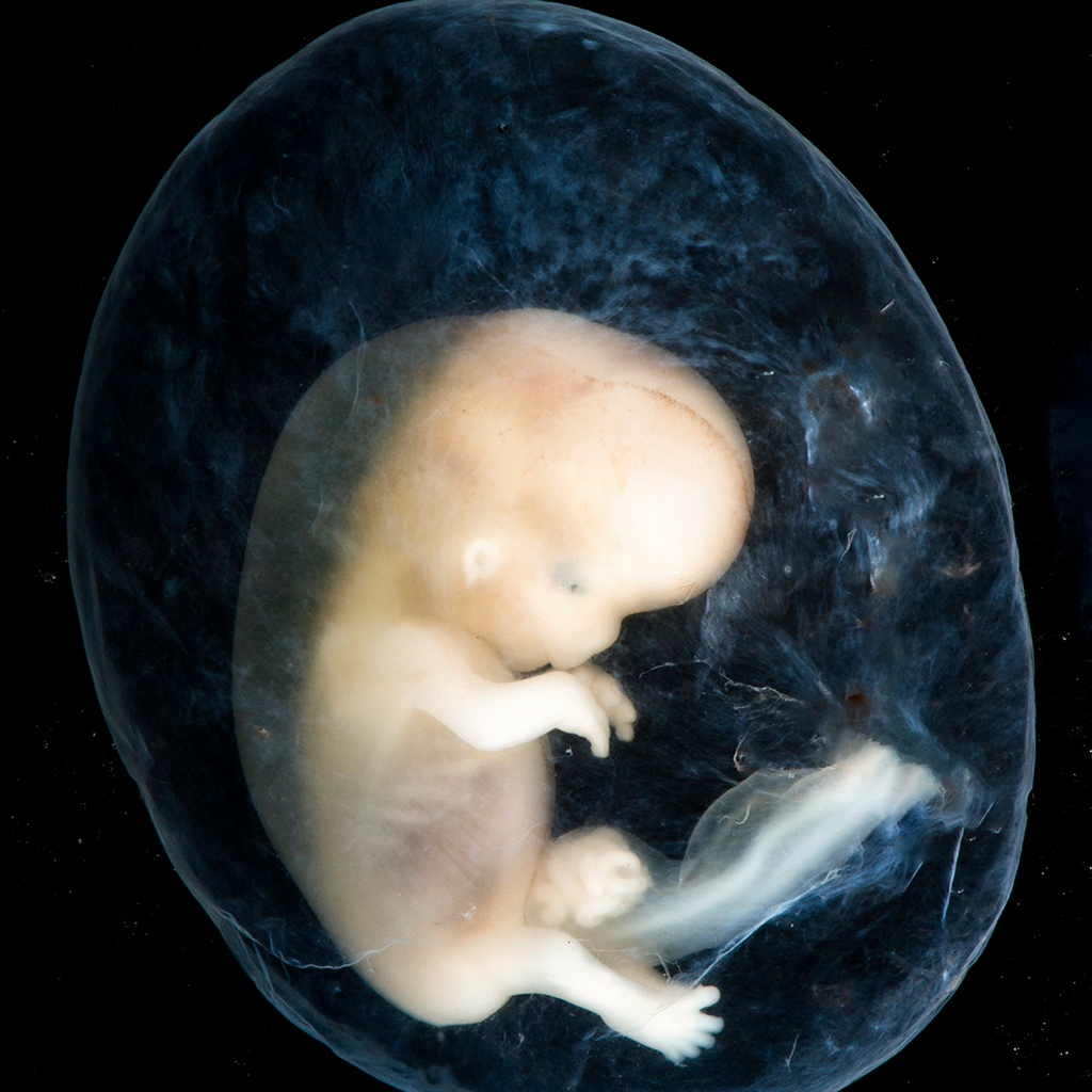
Embryo, 6- 7 weeks. After the first step, the cells begin to form a tiny human with a pine and a small, beating heart. The fetus gets nutrition to grow through the umbilical cord and soon develops arms, legs, and a face.
Embryo received with membranes intact. 1.6cm. EGA approximately 6-7 weeks from conception
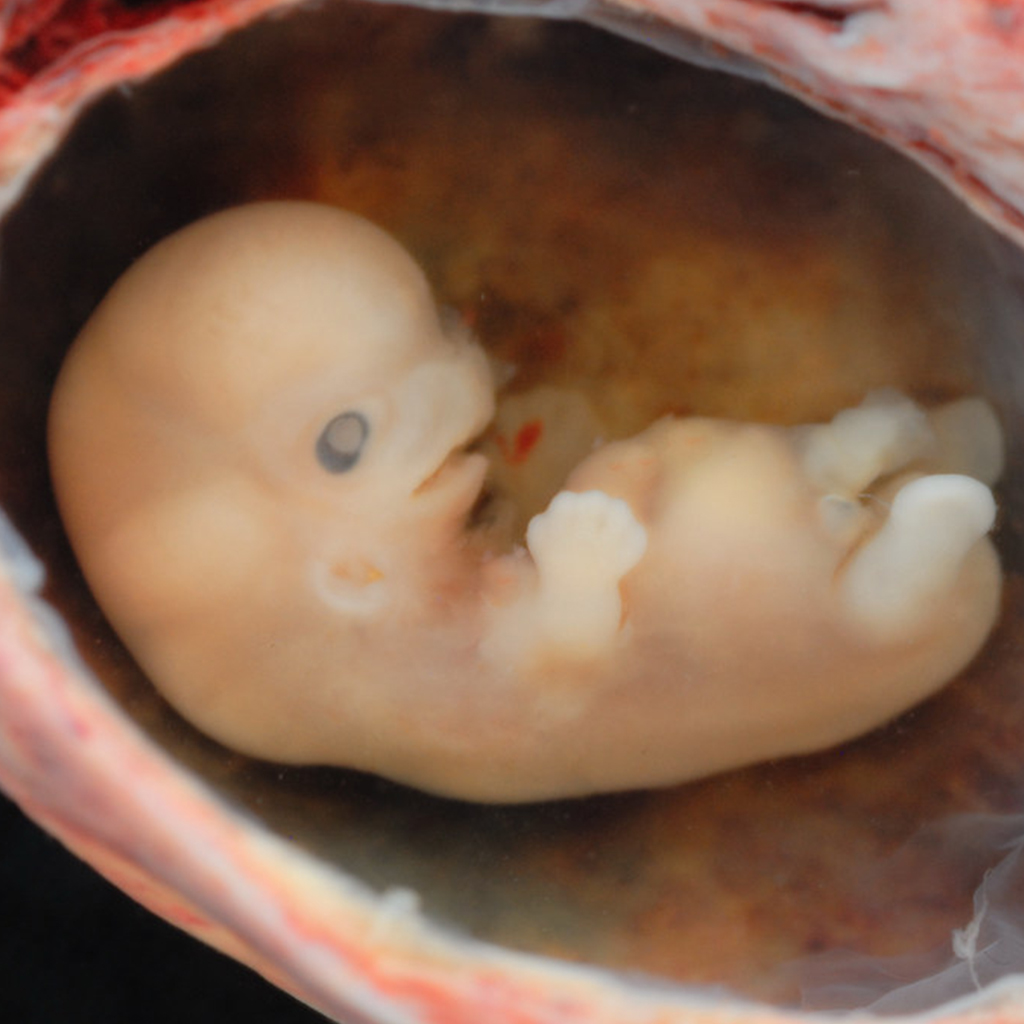
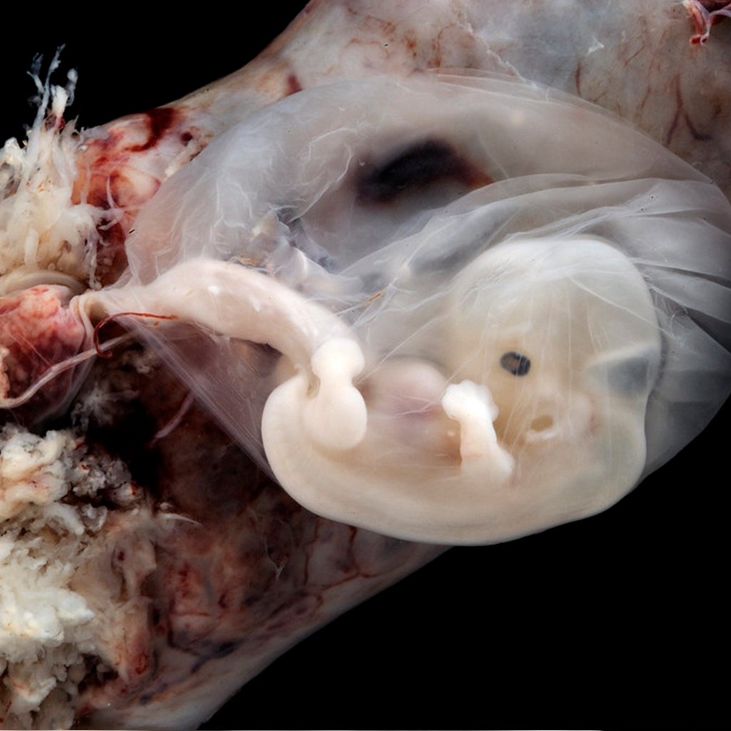
This is a 19mm embryo with an estimated gestational age of 6-7 weeks from conception, 8-9 weeks from LMP. Finger rays and brain hemispheres are among the features.
Embryo at 7 – 8 Weeks from conception
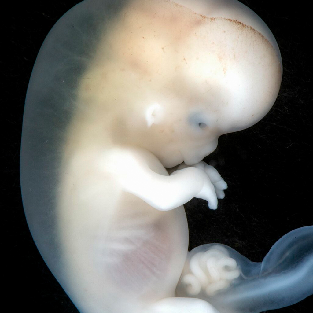
Embryo at approximately 8-10 weeks EGA from conception. This embryo still has its amniotic sac intact. The most notable finding is the extraembryonic coelom which contains loops of small bowel.
The most notable finding of this embryo is the loops of small intestine in an extraembryonic coelom. At 10 weeks these will return to the abdominal cavity.
Embryo week 9-10. Notable in the this fetus is an extraembryonic coelom in the umbilical cord. Intestinal loops are seen as a protrusion of orange material near the cord’s connection to the abdominal wall. At the end of the 11th week, these loops will be removed. The condition is known as an omphalocele if these do not return.
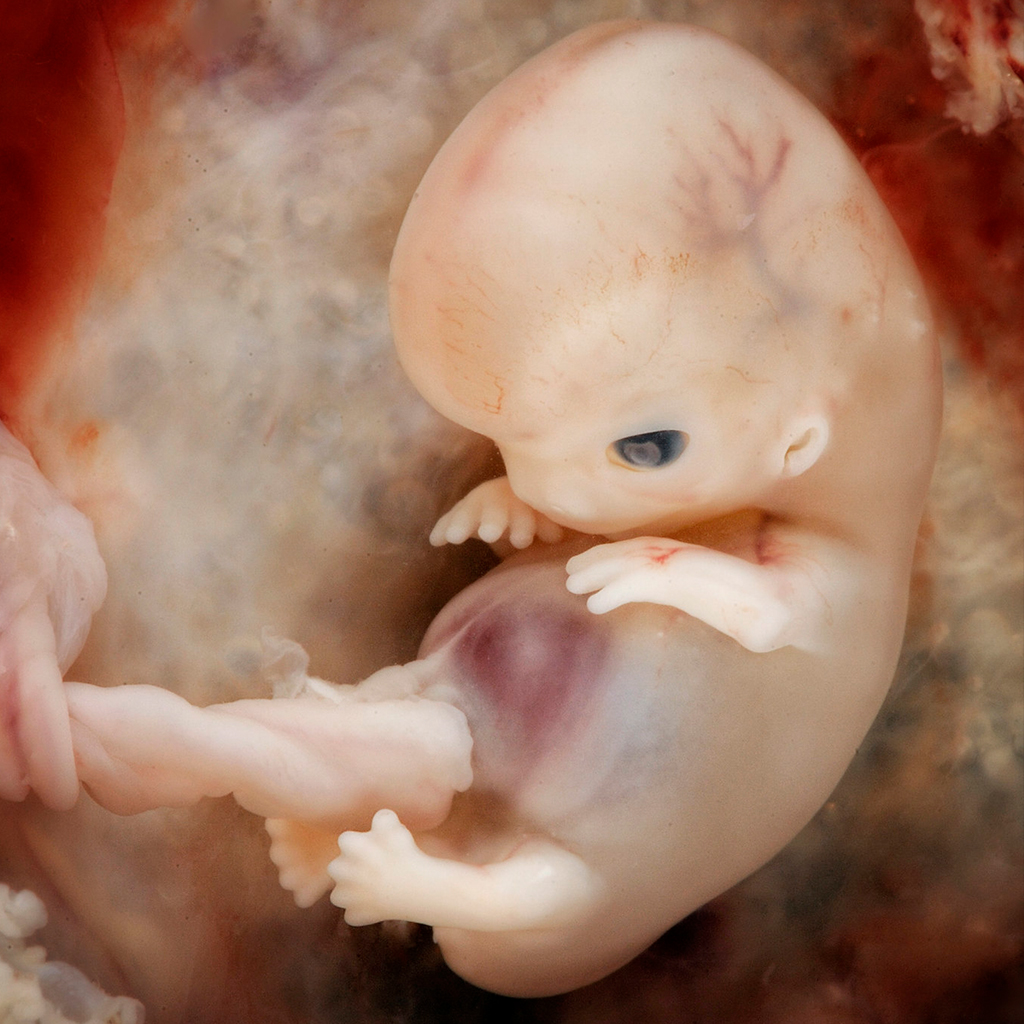
Fetus 10 – 12 weeks. CR 3.6cm. For comparison, the foot length is 0.6 cm, which is about the same length as the embryo in the photostream below at 3-4 weeks of development.
